
Functional MRI
How Does fMRI Measure Brain Function?fMRI uses MRI scanner to measure brain function. Click here to learn how MRI works.
Although there are other MRI methods to measure aspects of brain function, the method most commonly referred to in fMRI is the Blood Oxygenation Level Dependent (BOLD) method. BOLD uses a natural process. Hemoglobin in the blood carries oxygen to energy-hungry cells. The cells use the oxygen to produce ATP. The ATP molecules can be transported around the cell and used to drive biochemical reactions where and as needed.
 Active neurons in the brain require lots of energy and so they use more oxygen than resting neurons.
They get the increased oxygen from an increased blood flow. The mechanism by which the neurons call
for increased blood flow is unknown at this time. For some reason, also unknown, the blood flow
increase is far in excess of that needed by the neurons. The result is an increase in the blood flow
and an increase in the fraction of oxygenated hemoglobin in the blood.
Active neurons in the brain require lots of energy and so they use more oxygen than resting neurons.
They get the increased oxygen from an increased blood flow. The mechanism by which the neurons call
for increased blood flow is unknown at this time. For some reason, also unknown, the blood flow
increase is far in excess of that needed by the neurons. The result is an increase in the blood flow
and an increase in the fraction of oxygenated hemoglobin in the blood.
 Oxygenated and deoxygenated hemoglobin have different magnetic susceptibility. Brain tissue has a
susceptibility closer to that of oxygenated hemoglobin. Therefore, with more deoxygenated
hemoglobin in the blood, there is less disturbance in the magnetic fields and the spinning water
molecules exposed to the magnetic fields see less difference in spin velocity between positions.
As a result the net spin vector of the molecules stays larger and is measured as a larger MRI
signal. Thus, blood with a greater fraction of oxygenated hemoglobin sees a larger signal. This
corresponds with a greater fraction of oxygenated hemoglobin in active neurons.
Oxygenated and deoxygenated hemoglobin have different magnetic susceptibility. Brain tissue has a
susceptibility closer to that of oxygenated hemoglobin. Therefore, with more deoxygenated
hemoglobin in the blood, there is less disturbance in the magnetic fields and the spinning water
molecules exposed to the magnetic fields see less difference in spin velocity between positions.
As a result the net spin vector of the molecules stays larger and is measured as a larger MRI
signal. Thus, blood with a greater fraction of oxygenated hemoglobin sees a larger signal. This
corresponds with a greater fraction of oxygenated hemoglobin in active neurons.
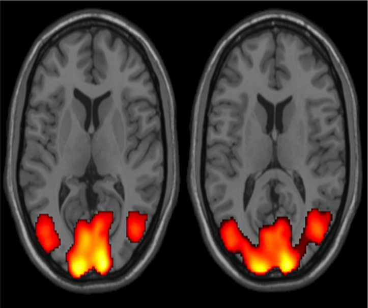
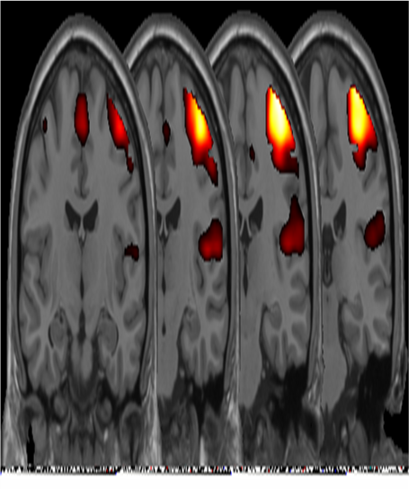
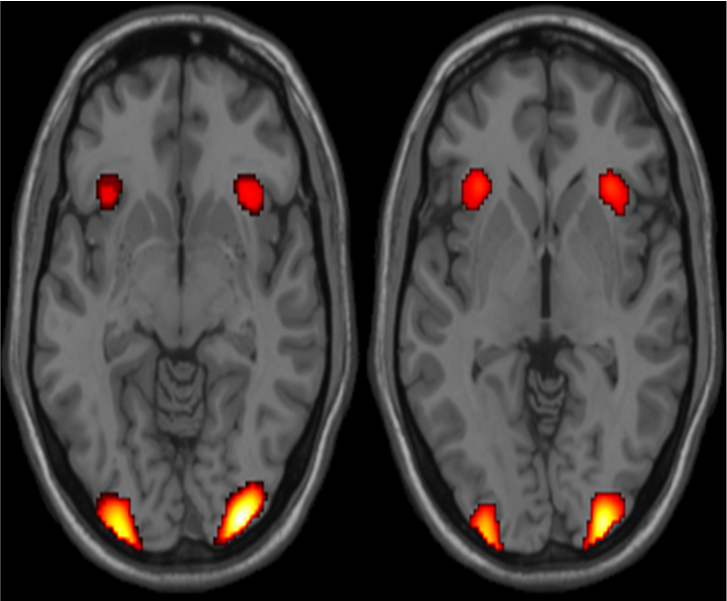 In the way described above, the neural activity in a location can be measured. fMRI is very good
at localizing the area compared to other techniques of brain imaging such as PET, SPECT or EEG, but
it is not as good as EEG at identifying the timing of neural activity. fMRI relies on the idea
that specific areas of the brain are called in as necessary to perform a cognitive task. It also
relies on people consistently using those specific areas.
In the way described above, the neural activity in a location can be measured. fMRI is very good
at localizing the area compared to other techniques of brain imaging such as PET, SPECT or EEG, but
it is not as good as EEG at identifying the timing of neural activity. fMRI relies on the idea
that specific areas of the brain are called in as necessary to perform a cognitive task. It also
relies on people consistently using those specific areas.
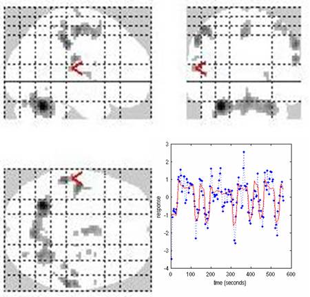
fMRI experiments use a cognitive task called a paradigm to generate specific neural activity. Paradigm is presented in a specific order in order to generate a timing pattern of (hopefully) consistent neural activity. The pattern of the paradigm is compared to the measured signal and areas of the brain that follow the pattern are identified as active in performing the paradigm.
 The change in MRI signal with oxygenated hemoglobin fraction is very small and is complicated by a
delayed response to the neural activity called the hemodynamic response. These things make it
difficult to detect the pattern even in areas that are active. We do as much as we can, but in the
end, the identification of active areas is a statistical decision and there is nothing certain.
The change in MRI signal with oxygenated hemoglobin fraction is very small and is complicated by a
delayed response to the neural activity called the hemodynamic response. These things make it
difficult to detect the pattern even in areas that are active. We do as much as we can, but in the
end, the identification of active areas is a statistical decision and there is nothing certain.
Read more: Wikipedia